











Medical Imaging Software Development
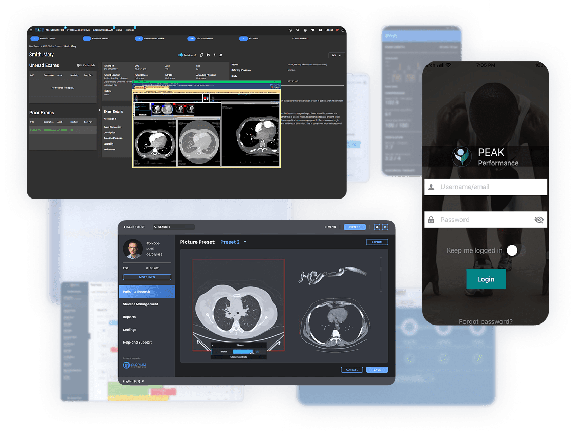

Smart Imaging Solutions for Better Patient Outcomes

- Universal image viewing
- Visualization features
- Full system compatibility

- Central image management
- Instant access to patient scans
- Scalable storage solutions

- Faster image interpretation
- Improved detection accuracy
- Priority case flagging

- High-def scan processing
- Faster imaging protocols
- User-friendly interface

- Real-time enhanced imaging
- Automated measurements
- Portable diagnostic options

- Rapid image processing
- Advanced 3D reconstruction
- Low-dose protocols

- 3D treatment planning
- Progress tracking tools
- CAD/CAM integration

- Millimeter-precise targeting
- Automated dose planning
- Real-time procedure tracking

- Clear cardiac imaging
- Automated calculations
- Standardized reporting
Imaging Software Services from Consulting to Support


Advanced Technologies Behind Medical Image Analysis Software


Turn Imaging Bottlenecks into Faster Diagnoses
- Calculate the cost of your project
- Discuss the timelines and roadmaps
- No commitment needed

Imaging Software Case Studies with Proven Results
- Irregular human anatomy made precise 3D reconstruction complex
- MRI, CT, and 3D ultrasound scans needed precise segmentation
- 2D-to-3D conversion had to be fast enough for surgical planning
- Imaging-to-3D model conversion now takes 30 minutes end to end
- DICOM viewer for STL files enabled detailed viewing of 3D models
- Creation of STL for 3D printing facilitated the production of physical 3D models
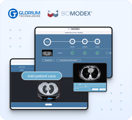
- Build a HIPAA-compliant platform from scratch after a previous vendor failure
- Create seamless integration between the physical scanner and the digital interface
- Develop medically accurate health tracking for chronic disease prevention
- 15+ vital metrics tracked, including body composition and disease markers
- Unity 3D interface for intuitive visualization of complex health data
- Wearable integration syncing with major fitness trackers for a 360-degree health picture
- Strict tech and budget criteria made senior Angular hiring hard
- One front-end center had to supply UI parts to many departments
- Growing in-house demand called for reusable, generic UI parts
- 2+ years of continuous Angular delivery alongside the core team
- Buttons, tables, grids, and sliders built for low-effort reuse
- Ongoing maintenance and upgrades kept the UI library current
Your Trusted Partner in Medical Imaging Software Development


Cost, Speed & Expertise: How Outsourcing Delivers More
 In-House Team
 Professional Outsourcing (Us)High ongoing salaries and infrastructurePredictable pricing, no HR overheadSlow ramp-up, training delays Immediate expert deploymentLimited to existing staff skills Specialized imaging developers on demandSteep learning curve for certifications ISO, FDA, and HIPAA-trained team
Image Analysis Software for Every Clinical Specialty








Tech Stack Behind Your Custom Solutions










How it Works
Your Custom Imaging Solution Is a Few Clicks Away


Can’t Find What You’re Looking For?


What our clients say about us







Success Stories










Who we are
About usGlorium Technologies is a full-cycle app & software development company which covers specific client business needs and manage them with the help of the best possible technology solutions.
Since 2010, we have been inventing digital breakthroughs, helping startups and businesses come out on top in their markets.

Why choose us




Certifications




Let's Connect!



Medical Imaging Software in 2026: What to Build, Integrate, and Plan For
Since the discovery of X-rays in the late 1800s, medical imaging has advanced significantly. Developments in artificial intelligence, cloud computing, and next-generation technologies are completely transforming the way we take, process, and interpret medical images. These technologies are improving patient outcomes and enabling quicker, more precise diagnoses. The market reflects this shift. Mordor Intelligence estimates the medical imaging software market at $8.75 billion in 2025 and expects it to reach $12.76 billion by 2030, a 7.8% CAGR. Demand for imaging also outpaces the people who read the scans: the Royal College of Radiologists’ 2025 census reports a 32% shortfall of clinical radiology consultants in the UK, with demand for complex imaging growing twice as fast as the workforce.
Automation, interoperability, and real-time processing are shaping the future of medical imaging software, from cloud-based imaging archives that are accessible from any location to AI-powered tumour detection. However, there are drawbacks to these developments as well, including issues with data security, legal compliance, and smooth clinical workflow integration. Successful implementation of this modern tech, compliance with regulatory standards, and security of patient data are possible through professional development. That’s why modern healthcare providers are turning to medical imaging product development consultants to ensure efficient integration and continuous improvement.
We’ve put together an article to explore the DICOM and PACS standards every imaging system relies on, the main advantages of AI, cloud platforms, edge computing, and new trends that are changing medical imaging, as well as what patients, developers, and healthcare professionals can anticipate in the years to come.
DICOM and PACS: The Standards Behind Every Medical Imaging Project
Every CT scanner, MRI machine, and ultrasound device in a modern hospital speaks the same language, and almost every image it produces lands in the same kind of system. That language is DICOM, and the system is PACS. Any medical imaging project, from a simple viewer to an AI diagnosis assistant, depends on handling both correctly.
What is DICOM?
DICOM (Digital Imaging and Communications in Medicine) is the international standard for storing, transmitting, and displaying medical images. NEMA publishes and maintains the standard, and ISO adopted it as ISO 12052. A DICOM file combines pixel data with a header of standardized tags, such as patient ID, modality, acquisition parameters, and study identifiers. Any compliant system can open the file and know exactly what the image shows.
For a development team, DICOM covers far more than a file format:
- DIMSE network services: C-STORE, C-FIND, C-MOVE, and C-GET send, query, and retrieve studies between medical imaging devices, PACS, and workstations
- DICOMweb: RESTful services (QIDO-RS for search, WADO-RS for retrieval, STOW-RS for storage) that let web and mobile apps work with imaging data over HTTPS
- Modality worklist (MWL) and MPPS: Send patient and order details from the RIS to the scanner and report back when the exam is complete
- Structured reports and segmentation objects: Store measurements, AI findings, and contours (DICOM SR, SEG, and RTSTRUCT) in a form other systems can read
- De-identification profiles: Rules in DICOM PS3.15 for removing patient identifiers before images go to medical research or AI training
Each DICOM-compliant device ships with a conformance statement that lists the services and data types it supports. Reading those statements at the start of a project tells your team what your DICOM imaging software must handle on day one.
What is PACS?
A Picture Archiving and Communication System (PACS) receives medical images from imaging equipment, stores them, and makes them available to radiologists and other healthcare professionals wherever they work. A typical PACS includes:
- Archive: Short- and long-term storage for medical images, often tiered between fast and low-cost storage
- Database and index: Tracks patients, studies, series, and images so users can find a scan in seconds
- Diagnostic viewer: Displays studies with hanging protocols, measurements, and 3D tools
- Integration layer: Connects the PACS to the RIS, EHR, reporting tools, and AI applications
Many healthcare organizations now pair or replace their PACS with a vendor-neutral archive (VNA). A VNA stores images from several departments and vendors in standard DICOM format, so switching viewers or PACS vendors no longer means migrating the whole archive. Cloud PACS goes a step further and moves storage and viewing to a hosted platform.
How DICOM, PACS, RIS, and EHR work together
Each system plays a distinct role in the medical imaging workflow:
| System | Main role | Standards used |
| Imaging modality (CT, MRI, ultrasound, X-ray) | Acquires medical images | DICOM, MWL, MPPS |
| PACS or VNA | Stores, indexes, and distributes images | DICOM, DICOMweb |
| Radiology information system (RIS) | Manages scheduling, orders, and reports | HL7 v2, DICOM MWL |
| Electronic health records (EHR) | Holds patient records and displays results | HL7 v2, FHIR |
| AI and image analysis tools | Analyze medical images and return findings | DICOM SR and SEG, DICOMweb, FHIR |
A typical exam follows this path. The physician places an order in the EHR, the RIS schedules the exam and sends a worklist entry to the scanner, and the scanner stores the images in the PACS. AI tools then analyze the study, and the radiologist’s report flows back to the EHR. A break at any step delays an accurate diagnosis, so integration testing deserves as much attention as feature development.
What to check before building DICOM and PACS software
- Conformance scope: Which DICOM services, SOP classes, and transfer syntaxes your software must support
- Integration targets: Which PACS, VNA, RIS, and EHR vendors and versions you need to connect to
- Data volume: Expected study sizes and daily volume, which shape storage, compression, and network design
- Data security: Encryption in transit (DICOM TLS), access controls, and audit logs that meet HIPAA requirements
- Validation data: Test studies from real modalities, not only sample files, to catch vendor-specific tag variations
AI Medical Image Analysis for Better Diagnostic Accuracy
Imagine a world in which potentially fatal situations are discovered in a matter of seconds rather than hours, where a dedicated assistant to radiologists never fails to notice minute tumours, subtle fractures, or early disease indicators. AI-powered medical image analysis has made that world a reality.
How AI sees what humans might miss
Traditional image review relies on the human eye, capable but prone to fatigue and variation. Due to these challenges, even professionals with years of experience scanning through medical imaging data may miss essential details.
AI delivers greater accuracy and changes the diagnostic process like so:
- Processing thousands of images in minutes: No coffee breaks needed
- Spotting patterns invisible to humans: Like micro-calcifications in mammograms or early ischemic strokes
- Learning from every scan it analyzes: It improves with each retraining on new, curated data
- Automating image segmentation: Deep learning algorithms outline organs, tumors, and vessels for surgical planning and radiation therapy planning
How does this translate into the real world? Recent studies have shown that AI-powered X-rays can detect early lung cancer and even reduce missed lung cancer diagnoses. Regulators have taken note. The FDA’s list of AI-enabled medical devices passed 1,500 authorizations by the end of March 2026, and about 76% of them are radiology tools, from stroke triage on CT scans to fracture detection on X-rays.
AI has slowly been augmenting specialists’ capabilities and acting as a second opinion on demand. But there are still some flaws. AI isn’t perfect; for example, it may flag harmless shadows as false positives. Some hospitals also struggle to connect AI tools with their PACS and RIS, especially when findings need to come back as DICOM Structured Reports or segmentation objects that radiologists can open in their usual viewer. However, these challenges can be avoided or mitigated with a professional medical imaging product development company. For example, at Glorium Technologies, we provide not only medical imaging software development services but also support and training for teams to be more comfortable working with advanced tech.
Cloud-Based Medical Imaging for Scalable Imaging Data
Imagine a radiologist in New York reviewing MRI scans from a rural clinic in real time—or a medical research team accessing thousands of anonymized MRIs and diagnostic information to train an AI model without moving a single hard drive. This is the power of cloud-based medical imaging, and it’s transforming how the healthcare industry manages visual data.
Bulky, on-premise servers are clunky. Cloud solutions offer:
- Anywhere access: Radiologists can securely review studies from home, a coffee shop, or across the globe
- Limitless storage: No more deleting old studies to make room for new ones
- Built-in disaster recovery: Automatic backups protect against data loss from hardware failures or natural disasters
- Cost efficiency: Hospitals pay only for the storage they use, avoiding massive upfront infrastructure investments
- Standards-based access: Cloud PACS and vendor-neutral archives expose studies through DICOMweb, so viewers and AI tools can retrieve medical images over secure HTTPS connections
What are the benefits of cloud tech in digital imaging?
Cloud-based imaging is now the functional foundation of contemporary healthcare systems, surpassing pilot projects and theoretical advantages. Early adopters of cloud solutions are already seeing measurable benefits, while late adopters run the risk of lagging behind in terms of productivity and the standard of patient care.
Healthcare organizations are realizing the transformative potential of cloud-based medical imaging, which is driving the shift. This transition is driven by three fundamental advantages that address long-standing challenges in medical imaging workflows, resource management, and technological innovation:
- Faster diagnoses through instant collaboration
- Better resource allocation (no more maintaining server rooms)
- Innovation at scale for AI and precision medicine
And there’s a new frontier in the medical imaging solution development industry: hybrid cloud-edge systems, where AI processes images locally while cloud storage provides secure sync and backup.
All three major cloud providers now offer managed DICOM services: AWS HealthImaging, the Google Cloud Healthcare API, and the DICOM service in Azure Health Data Services. Each one supports DICOMweb and built-in access controls, which shortens the path from prototype to production for cloud-based medical imaging products and protects data integrity during storage and transfer.
Edge Computing for Real-Time Image Processing
While cloud computing has transformed how we store and share medical images, edge computing is changing how we capture and analyze them in real time, especially in time-sensitive, on-the-go scenarios.
Every second counts when you’re dealing with critical care settings. Edge computing brings processing power directly to the imaging device—whether it’s a portable ultrasound, a portable MRI, or a mobile X-ray unit. This reduces the need to send data to the cloud. This means:
- Instant results: No lag time waiting for cloud processing
- Reliable performance: Works even with poor or no internet connectivity
- Enhanced privacy: Sensitive patient information can stay local when needed
For medical imaging applications development, this shift enables faster decision-making without sacrificing image quality or security.
Use cases where edge computing excels
| Healthcare Setting | Edge Computing Application | Benefits |
| Emergency Medicine | AI-assisted point-of-care ultrasound in ambulances | Speeds up triage by providing instant analysis during patient transport. |
| Portable CT scanners with stroke detection algorithms in ERs | Reduces time-to-diagnosis for critical conditions like strokes. | |
| Battlefield & Remote Diagnostics | Ruggedized imaging devices with onboard AI for field medics | Enables rapid injury assessment in resource-limited or combat environments. |
| Mobile ultrasound/X-ray applications for rural clinics | Provides advanced imaging capabilities without requiring a specialist on-site. | |
| Ambulatory & Primary Care | Compact imaging devices with edge AI for fracture/fluid detection | Helps GPs diagnose common conditions during routine visits. |
| Real-time image quality checks on portable systems | Ensures diagnostic-grade results even with lightweight equipment. |
Interoperability: Connecting Imaging Systems and EHRs
Modern medical imaging is a component of a complex digital health ecosystem, where smooth data exchange is essential. Interoperability, from AI-powered analytics to electronic health records, guarantees that physicians have the appropriate information at the appropriate time while preserving efficiency and security.
DICOM and PACS, covered earlier, handle the medical images themselves. The surrounding clinical context travels through other standards. HL7 v2 messages carry orders and results between electronic health records and the radiology information system (RIS), while HL7 FHIR resources such as ImagingStudy and DiagnosticReport let modern apps query imaging data next to the rest of the patient record. IHE integration profiles, such as Scheduled Workflow and XDS-I for cross-enterprise image sharing, define how all these pieces fit together.
Why is this important for medical imaging product development?
- AI tools are able to extract patient data in FHIR format and DICOM images for context-aware analysis
- By viewing studies directly within EHR interfaces, radiologists can minimize tab-switching
- Compliance with these standards is essential for medical device manufacturing and regulatory approval
Additionally, platforms like OHIF Viewer and 3D Slicer are transforming how teams interact with medical images:
| Platform | Key Role in Medical Imaging | Impact on Digital Health |
| OHIF Viewer | Web-based DICOM viewer with customizable user interface for hospitals & teleradiology | Enables remote second opinions without proprietary software |
| 3D Slicer | Advanced image fusion and segmentation for research/surgical planning | Supports precision medicine with multimodal data visualization |
| Orthanc | Lightweight open-source DICOM server with a REST API | Speeds up prototyping, research archives, and small-clinic PACS setups |
| MONAI | PyTorch-based framework for deep learning in medical imaging | Shortens the path from model training to clinical deployment |
These tools accelerate the software development process by providing ready-to-use frameworks for visualization, plug-and-play integrations, and modular design, which is essential for image analysis software development.
While still in the process of development, blockchain technology addresses two critical challenges:
- Patient data security
- Streamlined sharing
Blockchain solves these challenges for medical imaging product design and development by tracking every access to sensitive images and ensuring no single point of failure goes undetected; via smart contracts, it automates consent management for image sharing and gives patients control over who sees their data. This is essential for the peace of mind for worried cancer patients and for creating tamper-proof logs.
Emerging Trends That Shape the Future of Medical Imaging Technology
While we’re already implementing advanced tech that has proved essential for medical imaging software development, the future holds even more emerging technologies that may transform the healthcare industry. Let’s explore some of the trends that we, as a medical imaging software development company, are looking forward to:
3D/4D
Modern imaging is breaking free from 2D slices, offering clinicians dynamic, depth-aware perspectives:
- Surgical planning: Cardiologists navigate beating heart models in 4D (3D + time) to plan valve repairs
- Virtual biopsies: Radiologists “sample” tumors volumetrically in software before physical procedures
- Therapeutic monitoring: Track chemotherapy response via 3D tumor volume changes vs. crude diameter measurements
AR/VR
Augmented and virtual reality are moving from pilot projects to clinical staples:
| Application | How It Works | Impact |
| Surgical Navigation | AR overlays tumors/vessels onto real-world views during operations | Helps surgeons see tumor borders and vessels in context during resection |
| Medical Education | VR simulations let trainees “practice” on infinite virtual patients | Gives trainees repeatable practice cases without cadaver costs |
| Patient Consent | Interactive 3D models show procedures visually | Helps patients understand the procedure before they consent |
Federated learning
This breakthrough approach lets hospitals collaboratively train AI without sharing sensitive data:
- Local models train on each institution’s private datasets
- Only algorithm updates (not patient images) get aggregated
- Global model improves while data stays put
Foundation models and generative AI
Foundation models trained on millions of medical images are starting to reach clinical products. Instead of building one narrow model per finding, developers can fine-tune a single model for several tasks, such as triage, image segmentation, and draft report generation. Development teams still need curated imaging data, careful validation, and a regulatory plan for each intended use.
Better images alone won’t define the future; smarter, safer, and more integrated care is what’s needed. These trends require attention right now, regardless of whether you’re developing the next imaging AI or consulting on medical imaging software development.
Navigating FDA Clearance and Regulatory Compliance for Imaging Software
Building medical imaging software that works brilliantly in a test environment is one thing. Getting it legally approved for clinical use is another challenge entirely. Regulatory compliance isn’t a box you check at the end of development. It’s a framework that shapes every design decision, every line of code, and every testing protocol from day one.
Understanding software classification and its impact on your project
The FDA classifies medical imaging software as Software as a Medical Device (SaMD), and the regulatory pathway depends on the risk level. Class I devices (low risk, like simple medical image management software without diagnostic claims) may only require general controls. Class II devices (moderate risk, like AI-assisted detection tools) typically need a 510(k) submission demonstrating substantial equivalence to a predicate device. Class III is rare for imaging software.
Your software’s intended use statement drives this classification. A DICOM viewer used purely for reference falls into a different category than the same viewer enhanced with AI that flags suspicious nodules. Getting this classification right early prevents costly rework months into development.
For the European market, the EU Medical Device Regulation (MDR 2017/745) applies a similar risk-based classification, requiring CE marking and conformity assessment through a Notified Body for higher-risk classes.
IEC 62304: The software lifecycle standard you cannot skip
IEC 62304 defines the lifecycle processes required for medical device software development. It mandates specific activities based on your software’s safety classification (Class A, B, or C). For imaging software that informs diagnostic decisions, you’re typically looking at Class B or C, which means you need:
- Documented software development plans with defined milestones
- Detailed software architecture and design specifications
- Traceability matrices linking every requirement to its implementation and corresponding test case
- Formal verification and validation at each development phase
- A software maintenance plan covering the product’s entire commercial lifecycle
Skipping or retroactively applying these processes is a common and expensive mistake. Teams that treat IEC 62304 as an afterthought often face six to twelve months of additional documentation and re-testing before they can submit to regulators.
Design controls and quality management systems
Since February 2, 2026, the FDA’s Quality Management System Regulation (QMSR) under 21 CFR Part 820 incorporates ISO 13485:2016 by reference. One quality management system can now cover both the US and most international markets. Within this QMS, design controls create a structured loop: user needs feed into design inputs, which produce design outputs, which are verified against inputs and validated against user needs.
For medical imaging software, this means conducting formal usability studies with actual clinicians, performing risk analysis following ISO 14971 (identifying what happens if the software misidentifies a lesion or fails to load a scan), and documenting every design review and decision.
AI-specific requirements: PCCPs and the EU AI Act
Medical image analysis software that uses machine learning faces extra scrutiny. In December 2024, the FDA finalized guidance on Predetermined Change Control Plans (PCCPs). A PCCP describes planned model updates in the original submission, so manufacturers can roll them out later without filing a new 510(k).
The EU AI Act treats AI in medical devices that require Notified Body review as high-risk, with those obligations currently scheduled to apply from August 2, 2028. Planning for both early saves your development team from rebuilding documentation once the model is in clinical use.
Practical steps that keep your project on track
Working with a medical image analysis software development company experienced in regulated medical software simplifies this process considerably. Here’s what effective compliance integration looks like in practice:
- Risk management starts at kickoff: Preliminary hazard analysis during the planning phase identifies safety-critical features that need the most rigorous testing
- Regulatory strategy aligns with product strategy: Before writing code, define the intended use, target markets, and classification so development follows the correct pathway from the start
- Continuous documentation replaces last-minute scrambles: Design history files, test protocols, and traceability records are maintained in real time throughout development, not assembled retroactively
- Cybersecurity is baked in: Section 524B of the FD&C Act and the FDA’s premarket cybersecurity guidance require a Software Bill of Materials (SBOM), threat modeling, and a plan for addressing post-market vulnerabilities. For imaging software handling protected health information, this overlaps directly with HIPAA technical safeguards
Getting regulatory compliance right protects your investment, accelerates your time to market, and ensures your medical image analysis software development delivers solutions that can actually reach the clinicians who need them. It’s not overhead. It’s the foundation that makes everything else possible.
When to Hire a Medical Imaging Development Team
You can already see how quickly the industry is developing and adopting new technologies. It’s only a matter of time before the trends mentioned above become the norm. This means that the best time to take action and start working on medical imaging software development is now. But how do you know if you can do it in-house or need medical imaging product development consulting?
Here are a few factors that we’ve noticed prompt our clients to address us for professional help:
You’re building a new imaging product
When developing medical-grade software, knowing how to write code isn’t enough. This task requires deep DICOM/FHIR expertise for easy and compliant EHR/PACS integration. Additionally, you want to work with regulatory-aware development professionals to avoid legal risks and complications. On top of that, your medical imaging software development team needs to work on precision UX design, since new tools are always difficult to adopt, and your team members will need some confidence to start working with AI and cloud solutions.
A legacy system is holding you back
Legacy systems were helpful and essential, but today, some of them have become obsolete or outdated. With a clunky legacy system, you’re risking security issues from unsupported software, productivity drains from slow and non-interoperable tools, and miss opportunities. As a business in the healthcare sector – one of the most demanding and fast-paced industry – you’re responsible for adopting technology that supports your company’s growth. We recommend working with a medical imaging software development team to modernize your tech stack without disrupting clinical workflows and adding new features to your legacy system (if you’re not up for a new software implementation).
You want (or need) to implement AI
Many hospitals have started buying expensive AI tools and assistants that end up being unused because of poor integration, lack of clinician trust in “black box” algorithms, or unrealistic computational requirements. Nevertheless, adopting a new technology is always risky and requires attention from professionals. If you’re planning to implement AI into your existing systems (or want to build a new one), it’s high time to work with a professional medical imaging software development company.
You’re dealing with security or compliance gaps
HIPAA Journal’s analysis of HHS breach data counted more than 700 large healthcare data breaches in 2025, with hacking and IT incidents behind most of them. Because imaging archives hold huge volumes of sensitive patient data, you may need blockchain-secured audit trails for image access, zero-trust architectures for remote viewing, and compliance roadmaps for new FDA cybersecurity rules. All of this sounds too complicated and rarely do companies have in-house teams that possess all the necessary skills and expertise to make this happen. However, working with medical imaging software development professionals can help you avoid risky compliance gaps.
The cost of “DIY” vs. professional partnership
Finally, if you’re still debating whether to work with a professional medical imaging software development company or hire an in-house team, here’s a little table to help you understand the difference between these two.
| Scenario | DIY Approach Risks | Professional Solution Benefits |
| FDA submission | Rejections due to incomplete documentation | Submission-ready documentation built alongside the code |
| Cloud migration | Data corruption during transfer | Verified data integrity through checksums and study-level reconciliation |
| AI integration | Algorithm drift causing false positives | Continuous monitoring + recalibration services |
| PACS integration | Failed retrievals and missing DICOM tags discovered after go-live | Conformance testing against your PACS, VNA, and RIS before launch |
Start Building Medical Imaging Solutions with Glorium Technologies
At Glorium Technologies, we specialize in end-to-end medical imaging product development. We have professionals proficient in cutting-edge technology and real-world clinical needs. Whether you’re a healthcare provider, medical device manufacturer, or digital health startup, here’s how we drive success at every stage:
- We offer custom medical imaging software development
- Legacy system modernization
- DICOM and PACS integration, including DICOMweb APIs and PACS-to-VNA migration
- AI integration and deployment
- Security and compliance assurance
- Support and training for your team
We have 15+ years of experience in healthcare IT and provide a regulatory-first approach with agile development techniques. We’re sure you have hundreds of questions – and we’re always here to answer them. For more information and a free estimate of your project, request a discovery call with our representatives.

FAQs
Which algorithm is best in medical image processing?
There is no single “best” algorithm for medical image processing, as the optimal algorithm depends on the specific imaging modality and medical application. The choice of algorithm for medical image processing depends on factors such as image acquisition parameters, image noise, desired output, etc. Classic techniques such as edge detection and thresholding still work well for clearly defined structures, while deep learning algorithms such as U-Net handle complex image segmentation tasks.
Do you develop DICOM imaging software?
Yes, we do. We build DICOM viewers, routers, anonymizers, modality worklist integrations, and DICOMweb APIs, and we test each one against the DICOM conformance statements of your devices and PACS. Let us know your specific needs!
Which technologies do you use for building medical imaging software?
We use a range of technologies for medical imaging software development, including programming languages such as C++, Python, and Java, and libraries such as OpenCV, TensorFlow, and PyTorch, plus medical imaging toolkits such as ITK, VTK, DCMTK, pydicom, and MONAI.
What is the difference between DICOM and PACS?
DICOM is a standard: it defines how medical images and their metadata are formatted and exchanged. PACS is a system: it uses DICOM to receive, store, and distribute those images. A PACS can only exchange images with scanners and viewers that support the same parts of the DICOM standard.
How does a PACS work with a RIS and an EHR?
The RIS manages the radiology side of the exam: scheduling, worklists, and reports. The PACS stores the images, and the EHR holds the full patient record. HL7 v2 or FHIR messages link the RIS and EHR, while DICOM links the RIS, scanners, and PACS.
What is DICOMweb, and do I need it?
DICOMweb is the RESTful part of the DICOM standard. Web viewers, mobile apps, and cloud AI services use it to search, retrieve, and store images over HTTPS. If your medical imaging software runs in a browser or the cloud, DICOMweb support is almost always required.
Can you migrate images from a legacy PACS to a VNA or cloud PACS?
Yes. A typical migration includes a data audit, tag cleanup and normalization, phased transfer while the old system stays live, and study-by-study reconciliation. This approach protects data integrity and keeps radiologists working during the switch.
How do you de-identify medical images for AI training?
We follow the de-identification profiles in DICOM PS3.15. That means removing or replacing identifying tags, checking for private tags, and detecting text burned into the pixel data. Linked identifiers can be kept in a separate, access-controlled mapping when studies need to be re-identified later.
Which file formats besides DICOM does your imaging software support?
Research and AI pipelines often use NIfTI, NRRD, MINC, or Analyze formats, which are easier to process in bulk. We build converters between these formats and DICOM, so images can move from research tools back into clinical systems.