Advanced Medical Imaging: Exploring the Best Software Features


The rapid development of digital technologies in the last two decades has revolutionized just about every field of human activity, without any exception. The healthcare sector has obviously reaped a number of benefits from this digital revolution and software development.
In the 20th century, X-ray and ultrasound were the only two methods of medical imaging and diagnostics available to healthcare professionals. At the turn of the century, though, the choice became much broader — and today the list of advanced medical imaging tools wouldn’t even fit on one page.
So let’s start with the basics: what is medical imaging technology and what are the benefits of medical imaging at its current stage of tech development?
Content

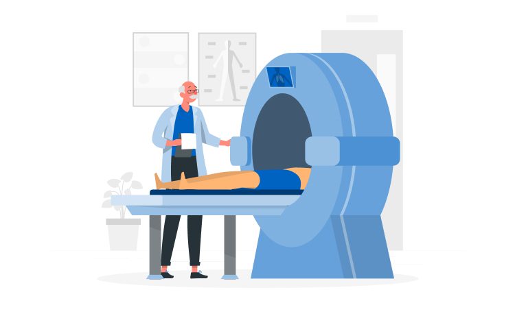
The most common examples of medical imaging are the good old X-ray, ultrasound, and MRI. We can refer to these methods as the first generation of medical imaging technology. These three tools have long-established benefits: they are relatively affordable (even for rural areas and poorer economies); and for 99% of all simple medical cases, first-generation medical imaging is universally available and instantly helps with both diagnostics and treatment decision-making.
The first generation of medical imaging technology has one significant drawback though — it is static. Thus, it would never be able to provide a complete picture of the case.
And the second generation of medical imaging tools — advanced medical imaging (often abbreviated as AMI in the academic & research environment) — closes exactly this loophole. Today, medical imaging technology is becoming dynamic, interactive, 3D, and even creative.
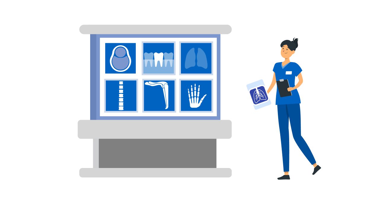
When you talk with healthcare professionals (especially administrators) about advanced medical imaging, one of the very first things they will mention is that medical imaging is a significant investment. And that’s a fact, medical imaging software may be costly, however, the tangible and intangible benefits from their implementation in medical practice definitely outweighs the related costs.
Clearly the number one benefit for all the patients, young and old. Also, contemporary methods of advanced medical imaging are often safer than the traditional X-ray, for example.
Advanced medical imaging development is nearly always instant. Moreover, the results it produces are not only immediate but also much more precise and complete. In a range of complex cases, such as cancers, immediate detection and diagnostics can be a real life-saver.
Software development for advanced medical imaging is indeed a serious expenditure item for hospitals and medical centers. On the other hand, for patients, insurance companies, and eventually for hospitals and clinics, the implementation of advanced medical imaging would mean lower costs for every single hospital stay. Simply because substituting invasive diagnostics & treatment methods with more advanced and non-invasive ones always leads to a saving effect. Take exploratory surgery as an example. Once you have advanced medical imaging as a readymade alternative, there is simply no need — in most cases — to perform exploratory surgeries as such.
In our observations, the most active users of medical imaging these days are surgeons and cardiologists. Although recently, advanced medical imaging also became popular among orthopedists and dentists. There is an easy, two-word explanation for this trend — 3D modeling.
Advanced medical imaging allows for laser-precise predictions of what will happen in case of complex intrusions into the human body.
In many complicated medical cases, surgical operations have undergone a real breakthrough (this is especially applicable to prosthetics). Experimenting during the course of an actual operation is highly risky and dangerously unpredictable. This is exactly where advanced imaging tools come in to help. With advanced medical imaging, surgeons can easily model the final outcome of an experimental operation, even if it has never been performed before.
For younger professionals, medical imaging also serves as the perfect learning tool. Using 3D simulations and predictive modeling tools, young doctors can reproduce the same operation multiple times in order to refine their surgical skills.
Medical imaging has not been developing in a vacuum, of course. Its progress is closely correlated with all other technological transformations in the sector. Thus, advanced medical imaging development has been put to immediate use, not only by doctors and nurses but also by hospital administrators.
Digitization of medical imaging tools now allows for their instant integration with other healthcare software solutions and hospital systems of record. From the customer feedback Glorium is currently collecting, most of today’s hospitals and other medical centers have already transferred their medical records to the cloud. Medical imaging tools have become an integral part of these cloud-based ecosystems. What does this mean on the level of provisioning daily medical services?
The benefits of medical imaging also include certain societal effects. Let’s be straightforward here, many patients (even the younger ones) still approach advanced diagnostic tools with a great deal of caution and fear. Even today, doctors often hear from patients that “X-rays shouldn’t be used more than twice a year.”
The earliest developments in the field of medical imaging might have indeed been questionable from the standpoint of health. However, in the last two decades, medical imaging science has been developing faster than ever before. Today, doctors and assisting personnel are already equipped with much better radiation protection gear, devices, and accessories. Thus, exposure to the so-called medical radiation is: 1) very precisely targeted; 2) greatly minimized. This is equally applicable to patients as well as the personnel administering medical imaging services.
In this area, much work is yet to be done to educate the general population about the current state of affairs in medical radiation use. Some patients, however, are already well aware that in today’s world, some office spaces might be far more dangerous for their health than the multiple diagnostics used in the administration of medical radiation.
Now that we’ve outlined what medical imaging technology is as well as the major benefits of medical imaging, let’s move on to specific examples. What’s under the hood of advanced medical imaging?

Also known as a helical X-ray or spiral CT scan. In very simplified terms, this is the traditional X-ray combined with a computer camera that is making a series of photos of the inside of our body.
The key benefit of a spiral scan is that it can produce multiple rotated images in a short time (thus, patients’ exposure to radiation is minimized).
Spiral CT scans are also much more detailed than regular X-ray images; thus, they can detect really tiny abnormalities at a much earlier stage.
In some cancer cases, spiral X-rays are not only used for diagnostics but also to check whether the targeted treatment is working.
DXR is a medical imaging technique based on obtaining sequential images using a flat-panel detector.
This method is mostly used for detecting lung diseases. Its key benefits are high image resolution and flexibility when it comes to body positioning. In the last two years, dynamic screening has been widely used to detect lung abnormalities after COVID-19.
In case of spinal abnormalities, surgeons often use both regular X-ray and DXR to compare the results and thus detect any causes for these abnormalities.

Most [relatively] healthy people have only seen this medical imaging technology in the movies. The full-body PET scanner is able to produce a full and precise 3D model of our body in as little as 20 seconds. Note that exposure to the human body, in this case, is not only very short-term but also less radioactive compared to most other techniques.
3D scanners are a huge step forward in medical diagnostics since the previous generation of these scanners (conventional, 2D ones) needed at least 20 minutes to scan the entire body.
Instantly working 3D scanners are a real life-saver for doctors working with small children (who either can’t lie calmly for an entire 20 minutes or are easily scared by lengthy interactions with an unusual device).
3D PET scanners have already demonstrated their high effectiveness in diagnosing atherosclerosis and other arterial-related diseases.
This is probably the most mathematical of all digital tools used in healthcare and medical analysis these days. Tracer kinetic modeling serves to visualize the time-and-space distribution of radiopharmaceuticals in the body. Thus, it is used not for diagnostics but in the course of treatment. Doctors even sometimes use visual graphs of tracer kinetic modeling to explain to their patients how the treatment is going and what processes are taking place in their body and what changes are happening on the molecular level (note that not all patients are open to this sort of explanation).

The logic that drives these newer ultrasound technologies is very much the same as with regular ultrasound — sound waves are sent out to produce an image according to their response from the “surface”. With 3D though, these sound waves are sent at multiple different angles simultaneously so as to compile a realistic, 3D image. And with 4D, the obtained images are then reproduced in a specific order. Thus, the 3D reflection looks like a short video clip.
3D/4D ultrasound software solutions have proved to be especially useful when doctors investigate inflammatory processes in the abdomen areas or observe how patients recover after bone injuries.
As the use of artificial intelligence is quickly spreading across all science-intensive sectors, healthcare and medical research are no exception.
The key benefit of using AI-based algorithms in CT (we haven’t listed CT in a separate category here), MRI, ultrasound, or endoscopy is the minimization of time needed to reproduce a full, highly detailed image.
AI-backed scanning equipment is not only quicker (thus, more efficient), in most cases, it also tends to be more portable. And this is a factor of critical importance for mobile and field hospitals.
Even for someone totally unfamiliar with healthcare and surgeries, these sorts of innovations do not require much of an explanation. VR simulations help design implants of a perfect, laser-precise size, shape, and angle. And then 3D printers help surgeons “grow” these implants from scratch. With 3D printing becoming widely popular in the healthcare sector, the costs of various implants have dwindled while the wait time (which used to be a literal real pain for patients) has fallen considerably. In some cases — only 24 hours.
As one of the most actively rising trends in today’s diagnostics, nuclear medicine imaging deserves a more detailed explanation.
This method of medical imaging is based on the detection of radiation tracers inside our bodies. In most cases, these radiation tracers are injected into veins (in very rare cases, they can be taken orally).
At first, the very idea sounds horrifying. However, in reality, the risks are minimal. First of all, the volume of radiation in a typical tracer intake is extremely low. As such, there won’t be any side effects from radiation after the procedure (unlike with chemo, for example). Moreover, nuclear medicine imaging does not use extra chemicals or artificial dyes (as was the case with the previous generation of scanners). This makes nuclear imaging perfect for people with severe allergies and/or weak immune systems.
Nuclear medicine imaging is mostly used in the diagnostics and treatment of cancers, and also in cases of blood disorders, thyroid diseases, and lung and kidney problems.
The great advantage of this method is that nuclear-based scanning shows how our organs are functioning in real time, and not simply how they look. This is the main reason why nuclear medicine imaging is mostly used not for diagnostics purposes but rather to check how a treatment is working inside the body.
During the last two years of the pandemic — despite all the budget constraints and medical investments being reprioritized — the amount of customer requests concerning customized software solutions for hospitals and medical research centers has been steadily growing, in our observations. An analysis of these requests allows us to forecast several trends that will be on the rise for medical software development over the coming years, and specifically in the area of advanced medical imaging development.

Two general IT-related trends will lead the pack throughout 2025 — cross-platform integrations and the hardline between cloud and on-premise solutions.
Describing cross-platform integrations would be easy in this particular case: doctors do not simply want to get faster diagnostics imagery and results — they want them instantly integrated into the system for patient records that they’re already using. Technically speaking, software solutions producing medical images must be 100% compatible with all other computer systems used in a hospital and other data storage systems in the first place.
Note that several different software vendors would offer software solutions within the same hospital. This does not necessarily mean that cross-platform integration isn’t possible due to a potential conflict of business interests. Software vendors these days are becoming more and more motivated by the capabilities of cross-platform collaboration. A range of vendors are already keeping their API libraries in open access, thus demonstrating their interest in cooperating with other vendors working with healthcare professionals.
However, the dilemma of “cloud vs on-premise solution” is a more tricky trend. As mentioned above, most of today’s hospitals (at least among our long-term clients and partners of our clients) have already begun storing their sensitive medical records in a secure cloud storage service. And one of the strong sides of having cloud-based storage is that it allows nearly instant data exchanges (with other doctors, research labs, etc.). At the same time, the issue of patient medical data privacy and how carefully that is managed under data sharing has to be considered.
Privacy issues, data security, and compliance regulations are the major reasons why hospitals are choosing on-premise systems for medical records. Is there a compromise to this dilemma? In fact, there is — hybrid software systems.
When applied to medical imaging data specifically, hybrid software allows comprehensive data exchanges on the one hand, while maintaining patient data privacy on the other.
During the last year, more than half of our current and prospective customers from the healthcare sector have demonstrated interest in such hybrid solutions — secure as closed, on-premise systems, but also readymade and open for collaboration with other hospital software systems as well.


